The Year’s Most Outstanding Science Visualizations
A juried competition honors photographs, illustrations, videos, posters, games and apps that marry art and science in an evocative way
![]()

First Place and People’s Choice, Photography: Biomineral Single Crystals. Credit: Pupa U. P. A. Gilbert and Christopher E. Killian; University of Wisconsin, Madison.
When Pupa U. P. A. Gilbert, a biophysicist at the University of Wisconsin, Madison, and her colleague Christopher E. Killian saw the scanning electron micrograph that they took of a sea urchin’s tooth, they were dumbstruck, says the journal Science. “I had never seen anything that beautiful,” Gilbert told the publication.
The individual crystals of calcite that form an urchin’s tooth are pointy, interlocking pieces; as the outermost crystals decay, others come to the surface, keeping the tooth sharp. In Photoshop, Gilbert added blues, greens and purples to the black-and-white image to differentiate the crystals. The resulting image calls to mind an eerie landscape in a Tim Burton movie.
Judges of the 2012 International Science & Engineering Visualization Challenge, a competition sponsored by Science and the National Science Foundation, as well as the public who voted online, were equally ecstatic about the SEM image. Enough so, in fact, that they selected the micrograph as the first place and people’s choice winner for the contest’s photography division.
The 10th annual Visualization Challenge received 215 entries across five categories—photography, illustration, posters and graphics, games and apps, and video. The submissions are judged based on visual impact, effective communication and originality.
And…drum roll, please. Here are some of the the recently announced winners:

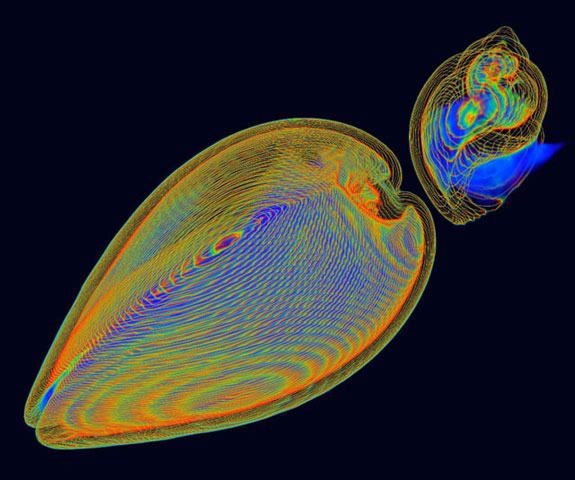
Honorable Mention, Photography: Self Defense. Credit: Kai-hung Fung, Pamela Youde Nethersole Eastern Hospital in Hong Kong.
Kai-hung Fung, a radiologist at Pamela Youde Nethersole Eastern Hospital in Hong Kong, captured this image of a clam shell (on the left) and a spiral-shaped sea snail shell (on the right) using a CT scanner. The image won honorable mention in the photography category. The multi-colored lines represent the contours in the shells. Fung told Science that he took into account “two sides of a coin” when making the image. “One side is factual information, wile the other side is artistic,” he told the journal.

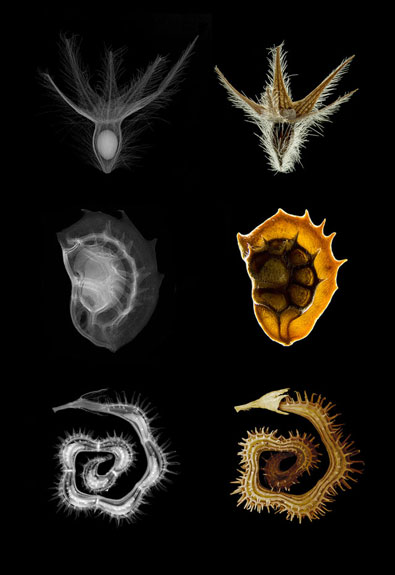
Honorable Mention, Photography: X-ray micro-radiography and microscopy of seeds. Credit: Viktor Sykora, Charles University; Jan Zemlicka, Frantisek Krejci, and Jan Jakubek, Czech Technical University.
Viktor Sykora, a biologist at Charles University in Prague, and researchers at the Czech Technical University submitted three miniscule (we’re talking three millimeters in diameter or less) seeds to high-resolution, high-contrast x-ray imaging (on the left) and microscopy (on the right). The above image also won honorable mention in the photography category.

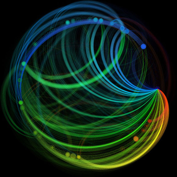
First Place, Illustration: Connectivity of a Cognitive Computer Based on the Macaque Brain. Credit: Emmett McQuinn, Theodore M. Wong, Pallab Datta, Myron D. Flickner, Raghavendra Singh, Steven K. Esser, Rathinakumar Appuswamy, William P. Risk, and Dharmendra S. Modha.
Earning him first prize in the illustration category, Emmett McQuinn, a hardware engineer at IBM, created this “wiring diagram” for a new kind of computer chip, based on the neural pathways in a macaque‘s brain.

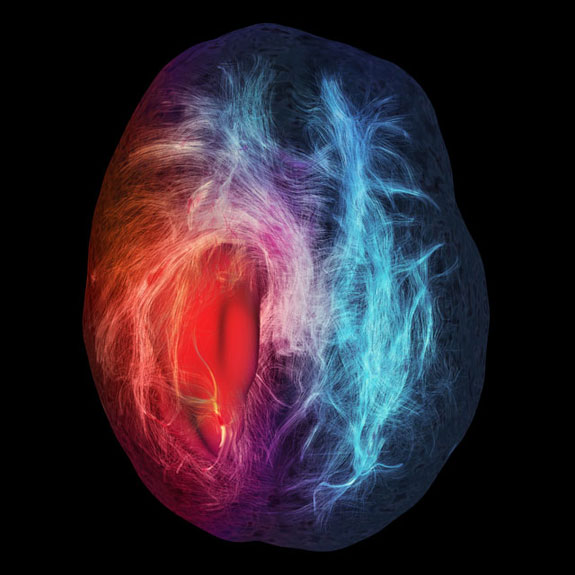
Honorable Mention and People’s Choice, Illustration: Cerebral Infiltration. Credit: Maxime Chamberland, David Fortin, and Maxime Descoteaux, Sherbrooke Connectivity Imaging Lab.
Maxime Chamberland, a computer science graduate student at the Sherbrooke Connectivity Imaging Lab in Canada, used magnetic resonance imaging (MRI) to capture this ominous image of a brain tumor. (The tumor is the solid red mass in the left side of the brain.) Science calls the image a “road map for neurosurgeons,” in that the red fibers are hot-button fibers that, if severed, could negatively impact the patient’s everyday functions, while blue fibers are nonthreatening. The image won honorable mention and was the people’s choice winner in the contest’s illustration category.
A team of researchers (Guillermo Marin, Fernando M. Cucchietti, Mariano Vázquez, Carlos Tripiana, Guillaume Houzeaux, Ruth Arís, Pierre Lafortune and Jazmin Aguado-Sierra) at the Barcelona Supercomputing Center produced this first-place and people’s-choice winning video, “Alya Red: A Computational Heart.” The film shows Alya Red, a realistic animation of a beating human heart that the scientists designed using MRI data.
“I was literally blown away,” Michael Reddy, a judge in the contest, told Science. “After the first time I watched the video, I thought, ‘I’ve just changed the way I thought about a heart.’”
Be sure to check out the other videos below, which received honorable mention in the contest:
Fertilization, by Thomas Brown, Stephen Boyd, Ron Collins, Mary Beth Clough, Kelvin Li, Erin Frederikson, Eric Small, Walid Aziz, Hoc Kho, Daniel Brown and Nobles Green Nucleus Medical Media
Observing the Coral Symbiome Using Laser Scanning Confocal Microscopy, by Christine E. Farrar, Zac H. Forsman, Ruth D. Gates, Jo-Ann C. Leong, and Robert J. Toonen, Hawaii Institute of Marine Biology, University of Hawaii, Manoa
Revealing Invisible Changes in the World, by Michael Rubinstein, Neal Wadhwa, Frédo Durand, William T. Freeman, Hao-Yu Wu, John Guttag, MIT; and Eugene Shih, Quanta Research Cambridge
For winners in the posters and graphics and games and apps categories, see the National Science Foundation’s special report on the International Science & Engineering Visualization Challenge.
/https://tf-cmsv2-smithsonianmag-media.s3.amazonaws.com/accounts/headshot/megan.png)
/https://tf-cmsv2-smithsonianmag-media.s3.amazonaws.com/accounts/headshot/megan.png)